Digital dentistry

From scan to smile, planned before the first incision.

Implant planning, surgical guides and smile design from your CBCT and scans, delivered remotely to clinics and labs in Egypt, the Gulf and Europe.

CBCT · DICOMBlueSkyPlanAtomica AIExocadDSD
Digital workflow

Every case follows the same four steps.

A digital workflow turns a CBCT scan into a precise surgical plan and a smile the patient can see in advance.

  1. Scan analysis

    Read the CBCT and DICOM data: bone, nerves, sinus and anatomy.

    CBCT · DICOM
  2. Implant planning

    Choose position, angle and size for each implant on the 3D model.

    BlueSkyPlan
  3. Surgical guide

    Design the guide that carries the plan to the chair.

    Atomica AI
  4. Smile design

    Design the final restoration and preview the result with the patient.

    Exocad · DSD

Implant surgical planning

Prosthetically driven plans from CBCT data, ready for the surgeon.

Digital Smile Design

Facial and dental analysis, smile mock-ups and before/after previews.

Surgical guide design

Tooth, bone and mucosa supported guides, designed for accuracy.

Crown & bridge design

Anatomic restorations designed in Exocad for lab and chairside work.

Backed by ten documented digital dentistry courses, five of them on surgical guide design. See all certificates

Cases

The result, and the plan behind it.

Step through each case, and click an image to view it full size.
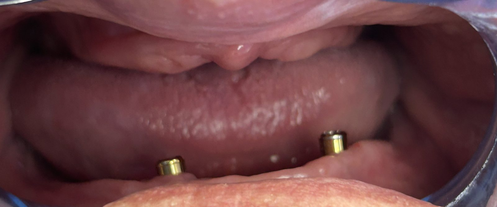
Clinical situation: two existing implants with attachments 1 / 7
Case 01 · Implant surgical planning

Lower implant overdenture: two implants added clear of the nerve

Upgrading a two-implant lower overdenture to four implants, with a digital plan that places each implant relative to the inferior alveolar canal and the mental foramen before surgery.

CBCT · DICOMNerve tracingAtomica AIGuided surgeryOverdenture
2 + 2Existing + planned implants
Ø4.1 × 10 · Ø4.1 × 8New implant sizes (mm)
0.2 mmCBCT voxel size
6 STLJaw and denture scans

Clinical situation

  • Edentulous patient wearing complete dentures
  • Lower overdenture retained by two interforaminal implants
  • Aim: more support and stability for the lower denture

Diagnostic data

  • CBCT (DICOM), 0.2 mm voxel
  • Jaw scans, plus denture scans with bite registrations
  • Scans registered to the CBCT in the planning software

Treatment plan

  • Inferior alveolar canal traced on both sides; mental foramina located
  • One implant per side, between the existing implants and the mental foramen
  • Axes aligned with the existing implants for a common path of insertion
  • Guide sleeves set for guided surgery

Planning outcome

  • Each implant fully within the bone on cross-sections
  • A safety margin kept between each implant and the nerve
  • Four-implant support instead of two: less denture rotation when chewing
  • Planned in the available bone, without grafting

Shared with the patient's consent, with identifying details removed. The plan is delivered to the treating surgeon, who keeps the clinical decision.
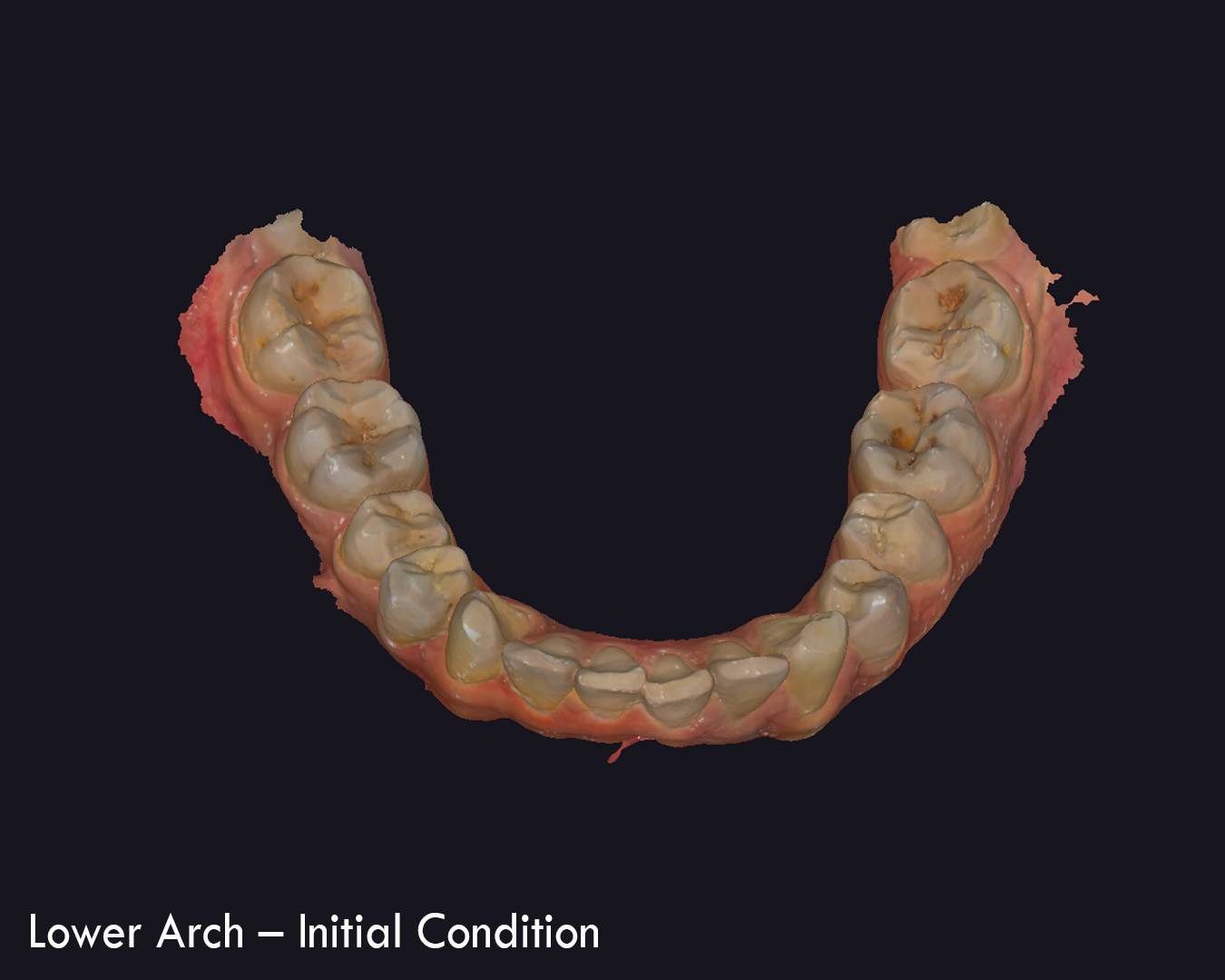
Lower arch, initial condition 1 / 4
Case 02 · Self case

Digital bite splint, lower arch

A bite splint I designed for my own occlusion: intraoral scans of both arches, the splint designed on the lower arch, and the new occlusal position checked digitally.

Bite splintDigital occlusionIntraoral scan

I am the patient in this case, shared with my own consent.

Case imageCBCT implant plan and surgical guide

CBCT plan and surgical guide

BlueSkyPlanAtomica AI
Case imageCrown and bridge restoration design

Restoration design

Exocad

Patient identities are always protected. Faces and personal data are covered or shared with consent.

Working together

Send your case in four steps.

I work remotely. You send the files, I send back a plan or design ready for your lab or your chair.

  1. Send the files

    Share CBCT (DICOM), intraoral scans (STL) and photos by Google Drive, WeTransfer or WhatsApp.

  2. Brief and quote

    Tell me what the case needs. I reply with my approach, price and delivery time.

  3. Design and review

    I share the plan or design for your review and adjust it until it is right.

  4. Delivery

    You receive files ready to print or mill, with a short report or case presentation.

Files I accept: DICOMSTLPLYOBJJPG / PNGMP4

Patient filesReceived through secure links, used only for your case, and deleted after delivery.

Professional scopeI work with licensed dentists and dental labs. Designs and plans are delivered to the treating clinician, who keeps the clinical decision and responsibility for treatment.

Ready to send your case?

Send the files on WhatsApp and I will reply with my approach and price.